Understanding Slit Lamp Parts and Functions: A Complete Guide for Eye Care Professionals
The slit lamp is a vital instrument in ophthalmology and optometry, providing a magnified, illuminated view of the eye’s anatomy. It allows eye care professionals to conduct detailed examinations of the anterior and posterior segments, which is crucial for diagnosing and managing various eye conditions. Knowing the components and their functions is essential for effective usage and accurate diagnosis.
1. Base and Support Arm
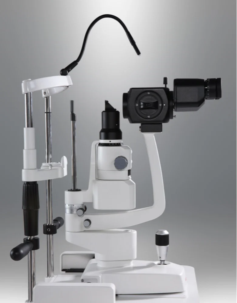
The base and support arm provide stability to the slit lamp, ensuring that the machine can be securely positioned for the examination. The support arm is adjustable, allowing practitioners to position it appropriately according to the patient’s height and positioning needs. Proper positioning is essential for obtaining a clear, focused image during the examination.
2. Headrest and Chinrest Assembly
This component helps maintain the patient’s head in a stable position. The headrest includes a forehead band, and the chinrest has an adjustable height to ensure comfort while the patient’s head remains still. This stability is key to obtaining consistent, detailed images, particularly during high-magnification examinations.
3. Biomicroscope
The biomicroscope is the core component that provides magnified, stereoscopic (3D) views of the eye. It allows professionals to inspect the eye’s structures layer by layer. Common features of the biomicroscope include:
- Eyepieces: Adjusted to the user’s interpupillary distance and prescription, if necessary.
- Objective Lens: Offers varying magnifications, usually between 6x to 40x.
- Magnification Changer: A rotating dial to switch magnification levels quickly for a more detailed view of specific areas.
The biomicroscope’s high magnification enables clinicians to examine subtle details within the eye, such as corneal cell layers, lens clarity, and retinal condition, essential for identifying diseases like cataracts or retinal detachment.
4. Light Source and Slit Mechanism
The slit mechanism is what makes this device a “slit” lamp. It includes a light source and a system of adjustable slits that control the width, height, and angle of the light beam. Key components include:
- Lamp Housing: Contains a halogen or LED light source.
- Slit Width and Height Adjusters: Control the size of the light beam, allowing for focused or diffuse illumination.
- Filters: Different filters—blue, green, red-free—help highlight specific eye structures, such as blood vessels or corneal abrasions.
By adjusting the angle and intensity of the light, the slit lamp can illuminate different layers of the eye. Narrow, intense beams are ideal for examining specific eye layers, while broader, diffused light is suitable for an overview of larger areas.
5. Joystick and Focusing Knobs
The joystick moves the slit lamp horizontally and vertically, while focusing knobs adjust the distance between the slit lamp and the patient’s eye. These adjustments allow eye care providers to focus precisely on the structure under examination and optimize clarity. Fine control over positioning ensures that structures like the cornea, iris, or retina are in focus and clearly visible.
6. Illumination Tower
The illumination tower houses the light source and slit mechanism, positioned at a specific angle to provide an oblique view of the eye’s anatomy. This angle allows clinicians to detect depth and curvature differences, such as corneal thickness or the depth of the anterior chamber.
The tower’s versatility makes it essential for procedures like gonioscopy, where indirect angles reveal the eye’s drainage angle, a key area in diagnosing glaucoma.
7. Filters and Lenses
Different filters serve specific diagnostic functions:
- Red-Free Filter: Enhances blood vessel contrast, helping detect vascular issues.
- Cobalt Blue Filter: Used with fluorescein dye, it highlights corneal abrasions, ulcers, and foreign bodies.
- Neutral Density Filter: Reduces light intensity for sensitive patients or when examining light-sensitive structures.
Some slit lamps also include additional lenses for specialized imaging, such as fundus lenses, which enable visualization of the posterior eye structures.
Using the Slit Lamp for Comprehensive Eye Examinations
Understanding each part’s functionality helps practitioners conduct more effective exams and produce reliable diagnostic images. Proper setup, including adjustments to the biomicroscope, light, and slit mechanism, is essential for thorough examination and accurate diagnosis. The slit lamp’s versatility, from wide-field imaging to detailed layer-by-layer examination, underscores its essential role in eye care.
Maintenance Tips for Optimal Performance
Regular maintenance is critical for extending the lifespan of a slit lamp and ensuring accurate results. This includes:
- Cleaning the lenses and mirrors with appropriate materials to prevent dust buildup.
- Regularly checking and replacing the light source to maintain consistent illumination.
- Adjusting and tightening mechanical parts to avoid instability during exams.
In summary, the slit lamp remains a cornerstone of eye examinations, allowing eye care professionals to diagnose and monitor a wide range of eye conditions effectively. By mastering each part and its function, practitioners can enhance the quality of care they provide, benefiting patients through detailed, accurate assessments of their eye health.



Hairstyles
hello there and thanks for your information ?I抳e definitely picked up anything new from right here. I did then again experience a few technical issues the use of this web site, since I skilled to reload the website a lot of times previous to I may get it to load properly. I were considering if your hosting is OK? Now not that I am complaining, however slow loading cases times will very frequently impact your placement in google and could injury your high quality score if ads and ***********|advertising|advertising|advertising and *********** with Adwords. Anyway I am including this RSS to my email and could look out for a lot extra of your respective interesting content. Make sure you replace this again very soon..