How Computed Radiography Works: Understanding the Imaging Process
Computed Radiography (CR) is a widely used imaging technology that provides a digital alternative to traditional film-based radiography, improving diagnostic accuracy, efficiency, and patient outcomes. This guide explains how CR works, from capturing the initial image to generating high-quality digital results, making it an essential tool in modern diagnostic imaging.

Traumatologist female doctor reviewing an x-ray of a patient’s hands. Skeleton bone disease exam and medic aid.
1. What is Computed Radiography?
Computed Radiography is an imaging technique that converts X-ray images into digital files without using traditional film. Unlike Digital Radiography (DR), which captures images directly via electronic sensors, CR uses photostimulable phosphor plates (PSPs) to capture and process images.
2. Key Components of Computed Radiography
- Phosphor Plates (PSPs): CR uses reusable PSPs, which are coated with a special phosphor material that captures the X-ray image.
- Image Reader: The image reader scans the PSP to convert the stored image into a digital format.
- X-Ray Source: Like traditional X-ray systems, CR uses an X-ray source to create images of the body’s internal structures.
- Computer System: The CR image reader connects to a computer where the digital image can be enhanced, reviewed, and stored.
3. The CR Imaging Process: Step-by-Step
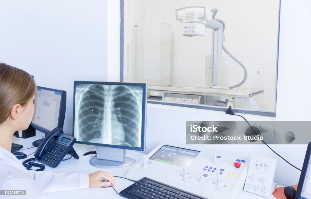
Step 1: Positioning and Exposure
The patient is positioned based on the imaging requirements, and the X-ray source directs beams toward the area of interest. The X-rays penetrate different tissues and are absorbed variably, creating an image on the phosphor plate placed behind the patient.
Step 2: Phosphor Plate Image Capture
The phosphor plate absorbs and stores the energy from the X-ray exposure. Areas with more X-ray absorption (like dense bones) appear darker, while areas with less absorption (like soft tissues) appear lighter. This contrast creates the foundational image on the plate, similar to how traditional film works.
Step 3: Scanning the Phosphor Plate
The exposed PSP is then removed from the cassette and inserted into a CR image reader, where it undergoes a laser scanning process. During this scan, the laser stimulates the phosphor on the plate, releasing stored energy in the form of light.
Step 4: Light Detection and Conversion
The light emitted from the phosphor plate during the laser scan is then captured by a photomultiplier tube (PMT) or a similar light sensor in the image reader. This light signal is amplified and converted into an electronic signal, creating a digital representation of the original X-ray image.
Step 5: Digital Image Processing
The electronic data from the PSP is processed into a digital image file. The CR system software adjusts brightness, contrast, and sharpness to enhance diagnostic quality. Technicians can further refine these settings if needed.
Step 6: Image Storage and Review
The finalized digital image is saved in the Picture Archiving and Communication System (PACS) for easy access, retrieval, and sharing among medical teams. CR images can be reviewed on a computer screen, allowing radiologists to zoom in, annotate, and make accurate diagnoses.
4. Advantages of Computed Radiography
- High-Quality Digital Imaging: CR offers high-resolution images that are ideal for a variety of diagnostic applications.
- Improved Efficiency and Speed: Unlike traditional film, CR images are processed within seconds, reducing the wait time for patients and clinicians.
- Cost-Effectiveness: Since PSPs are reusable, CR is a cost-effective option for hospitals that want to digitize their radiology departments without the immediate expense of DR systems.
- Digital Storage and Sharing: CR images are easily stored, retrieved, and shared digitally, supporting collaborative diagnosis and streamlined patient care.
5. Limitations and Considerations
- Maintenance and Calibration: PSPs and image readers require regular maintenance for optimal performance and accuracy.
- Image Quality Factors: CR images may suffer from minor loss of detail compared to DR systems. However, image enhancement options often compensate for this.
- Radiation Exposure: CR does reduce the radiation dose compared to traditional film, but it may still be higher than that of DR systems, making radiation management essential.
6. Comparing Computed Radiography with Digital Radiography
- Image Quality: DR often has higher image quality due to direct capture, but CR remains highly effective for most clinical needs.
- Cost and Implementation: CR is more affordable for facilities transitioning from analog to digital, whereas DR requires more upfront investment.
- Flexibility: CR is more adaptable in setups with existing analog equipment, allowing a gradual transition to full digital imaging.
7. Future of Computed Radiography
The future of CR lies in ongoing advancements, such as improving PSP sensitivity, enhancing image processing algorithms, and integrating artificial intelligence (AI) for image analysis. These innovations may bridge the gap between CR and DR in terms of image quality and speed, ensuring that CR remains a viable and efficient imaging solution.
Conclusion
Computed Radiography plays an essential role in healthcare by bridging the gap between traditional film and fully digital radiography. Its efficiency, cost-effectiveness, and diagnostic capabilities make it a valuable asset, especially for healthcare facilities looking to improve imaging quality without significant infrastructure changes. Understanding how CR works can help healthcare providers optimize its use for more accurate, timely patient diagnoses and enhanced care.

Leave a Reply